
If you've been living with jaw pain that won't settle down, facial tenderness that keeps moving around, or headaches that never quite match the dental X-rays you've already had, you're not alone. People often arrive at a specialty clinic after trying pain relievers, a night guard, and one round of imaging after another, only to hear that nothing “serious” shows up. That gap between how bad the pain feels and what the pictures show is exactly where a 3-phase bone scan can become useful.

A 3-phase bone scan looks at blood flow, soft-tissue pooling, and bone turnover in sequence, which gives clinicians a way to see active change instead of just anatomy. That matters in facial pain because the source may be a joint, a bone, or surrounding tissue, and those problems don't always look obvious on a standard dental film or even a CT scan. If you're also sorting through other causes of facial discomfort, a focused resource like facial skin restoration options can help you think broadly about orofacial symptoms while your medical team narrows the diagnosis. For a plain-language overview of causes, you may also find what causes facial pain helpful.
Practical rule: if the pain is persistent and the usual images are unrevealing, clinicians often need a test that shows activity, not just structure.
When Jaw Pain Needs More Than an X-Ray
A patient with TMJ pain often describes a familiar pattern. The jaw aches near the ear, chewing feels tiring, and the face may feel sore or swollen by the end of the day. Standard dental X-rays can look normal even when the person is clearly hurting, because X-rays mainly show shape and density, not whether a joint or bone is actively inflamed.
That's where the 3-phase bone scan earns its place. Major clinical references describe bone scans as useful when the cause of bone pain is unclear or when infection or occult injury is suspected, because nuclear imaging can detect abnormal tracer uptake before obvious anatomic change appears historical role and diagnostic value. In a TMJ setting, that means the clinician isn't just asking, “Does the joint look worn?” The question becomes, “Is the joint actively reacting right now?”
A scan like this is also different from a plain structural study because it follows physiology over time. The early images show blood supply, the middle images show soft-tissue pooling, and the delayed images show bone activity. That sequence helps separate a joint problem from a nearby soft-tissue issue, which is often the primary challenge in facial pain.
If you've already been told that “nothing major shows up,” this test can feel reassuring because it doesn't rely on a single still image. It asks a more useful question for pain care, which is whether the tissue is behaving like an active source of symptoms.
How the Three Phases Work Together
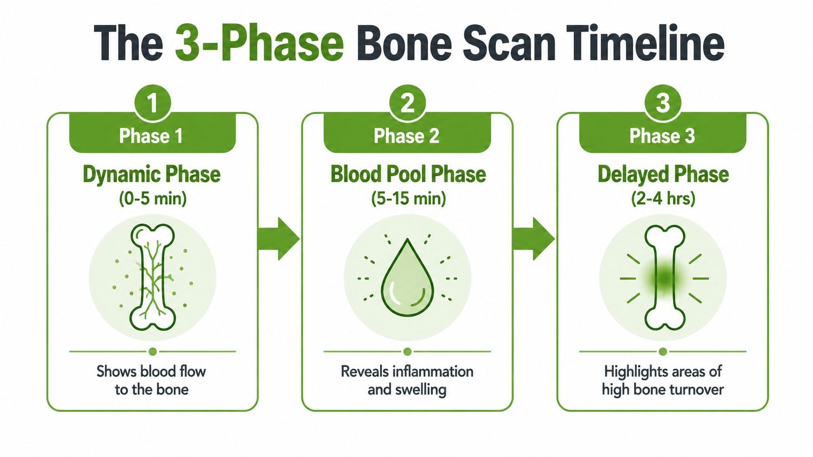
A 3-phase bone scan is a dynamic nuclear medicine study built around three timed image sets, and the timing is the whole point. The tracer is typically a technetium-99m compound, and the sequence starts during injection, continues almost immediately afterward, then finishes with delayed images hours later technical overview.
Phase 1 shows blood flow
The first phase is the dynamic flow phase. The camera records images at rapid intervals, and standard references describe 1-second intervals for 60 seconds during this early capture timing details. In plain language, this is the scan's way of watching how quickly tracer reaches the area.
Phase 2 shows blood pooling in soft tissue
The second phase is the blood-pool phase, taken right after the angiogram and within minutes. The scan shows whether the region is holding extra tracer in the soft tissues, which often points to local inflammation or swelling phase definition. In a facial pain workup, that can help clinicians tell whether the issue is more joint-centered or more soft-tissue driven.
Phase 3 shows the bone's delayed response
The third phase is the delayed skeletal image, usually acquired about 2 to 4 hours after injection, with patient-facing guidance often scheduling it around 3 to 4 hours later timing details. This phase highlights areas of higher bone turnover, which is why the scan can reveal early pathology before a structural image changes.
The scan reads like a timeline. First circulation, then soft-tissue response, then bone response. That order is what gives the test its clinical value.
Conditions a 3-Phase Bone Scan Can Detect
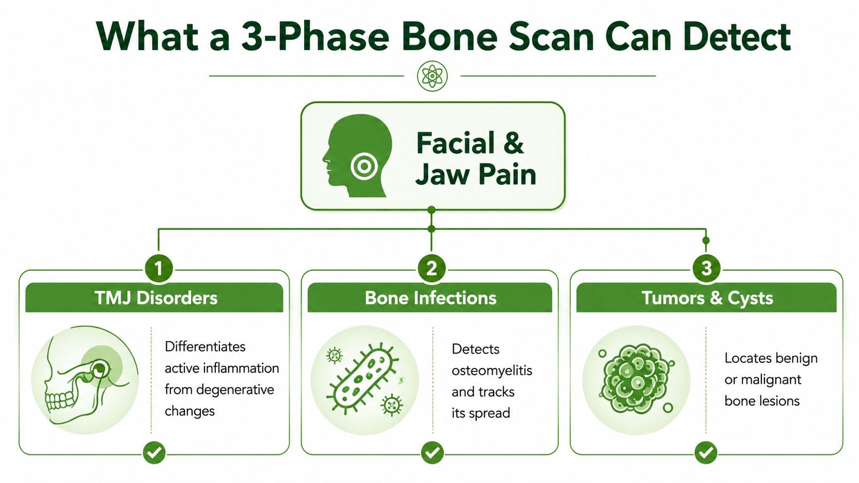
For TMJ and facial pain patients, the scan is most helpful when the clinician suspects active inflammation, bone infection, or occult injury. It is especially valuable because the result depends on a pattern across phases, not just one bright spot on a final image.
Why infection shows a recognizable pattern
In suspected osteomyelitis, early hyperemia appears as increased regional perfusion on the flow and blood-pool images, and if infection is present, that is followed by focal increased tracer uptake on delayed skeletal images classic three-phase pattern. That cause-and-effect pattern helps differentiate osteomyelitis from cellulitis, which may involve the soft tissue without the same delayed bone signature differentiation reference.
Why the scan can matter early
Nuclear medicine references note that the exam can become positive within 24 hours of symptom onset for osteomyelitis early positivity. That doesn't mean every new ache needs a scan. It does mean the test can sometimes detect activity early enough to guide treatment before a problem becomes harder to manage.
Why sensitivity still comes with limits
The scan is sensitive to bone turnover, which helps with problems such as suspected osteomyelitis, occult fracture, complex regional pain syndrome, and hardware-related complications clinical usefulness. The tradeoff is specificity, because healing fractures and other bone conditions can also look active on delayed images. In other words, a positive scan tells the team that something is biologically active, but it still has to be interpreted with the history, exam, and other imaging.
Preparing for Your Scan and What to Expect
Most patients are surprised by how straightforward the appointment feels. The test starts with a tracer injection, usually a technetium-99m-labeled compound, and then the imaging team captures the phases in sequence while you rest and wait between scans radiotracer family. For facial and TMJ studies, the camera is positioned near the jaw and face, so the technologist can focus on the region that matters most.
What the visit usually feels like
You'll likely spend some time on the imaging table lying still while the camera moves or stays positioned over the area. The immediate phase is quick, but the delayed phase means the whole appointment stretches over hours because the tracer needs time to reach the bone and settle into areas of higher turnover. That waiting period is part of the test, not a delay caused by a problem.
Why less imaging can still be enough
Evidence published in AJR found that routine delayed whole-body imaging after a directed 3-phase study did not improve diagnostic yield or alter patient care for most indications, so it was considered an unnecessary resource use AJR finding. In practical terms, many focused exams don't need a full extra survey if the clinical question is already local and specific.
A few basics help the day go smoothly:
- Wear simple clothing, because you may want to avoid metal around the face and chest.
- Tell the technologist about pregnancy or allergies, since those details matter for safe imaging.
- Plan for waiting time, because the delayed phase is built into the study.
- Ask about your local protocol, since patient instructions can differ by clinic and by the exact area being scanned.
The most stressful part is usually the uncertainty beforehand. Once the steps are explained, the test is often more predictable than people expect.
Comparing Imaging Options for Facial Pain
Patients often assume every advanced scan answers the same question, but each modality looks at something different. A 3-phase bone scan is best when the team wants to know whether tissue is actively inflamed or metabolically stressed. Other scans can be better for anatomy, joint mechanics, or soft tissue detail.
| Imaging Type | What It Shows | Best For | Limitation |
|---|---|---|---|
| 3-phase bone scan | Blood flow, soft-tissue pooling, delayed bone activity | Suspected active inflammation, infection, occult injury, joint involvement | Less specific, because several conditions can increase uptake |
| CBCT | Fine bony anatomy and joint structure | Dental and bony detail, alignment, structural changes | Does not show blood flow or active inflammation |
| MRI | Soft tissue, disc position, joint effusion | Disc problems, soft tissue inflammation, joint fluid | Less focused on early bone metabolic change |
| SPECT/CT | Functional uptake plus anatomical localization | Pinpointing where uptake sits in relation to structure | More specialized, and interpretation depends on the combined study |
CBCT and MRI often answer different questions than a bone scan. CBCT is excellent for structural detail, while MRI is stronger for soft tissue and the TMJ disc, so those studies are often complementary rather than competing. If the goal is to localize biologic activity and then match it to anatomy, SPECT/CT can add another layer of precision, especially in complex cases.
For patients who want a specialty evaluation of that bigger picture, tmj specialist Raleigh NC is a useful search phrase to understand how clinics organize these kinds of diagnostic decisions. The key idea is simple. The right scan depends on the question being asked, not on which test sounds most advanced.
How Scan Results Shape Your Treatment Plan
A scan is useful only if it changes what the clinician does next. In a TMJ or facial pain workup, evidence of active joint inflammation can support a plan that stays focused on the joint itself, while a scan with no convincing bone involvement can push the team to look harder at muscular tension, posture, airway strain, or other non-bony sources of pain.
That distinction matters because treatment choices can change with the diagnosis. If the problem looks joint-based, a clinician may consider Prolotherapy or Platelet-Rich Fibrin injections, and patients often ask about orthobiologic therapy when they want a non-surgical path. If the scan does not show bone involvement, care may shift toward oral appliances, cold laser therapy, or therapies aimed at muscle coordination and breathing patterns instead.
A good scan doesn't just name a problem. It helps prevent the wrong treatment from being repeated.
That's especially important in facial pain, where symptoms can overlap. One person's pain may start in the joint, while another's comes from the muscles that stabilize the jaw. A 3-phase study helps separate those possibilities so the plan is based on the tissue that's active.
If jaw pain, facial aching, or unexplained TMJ symptoms have stayed unresolved despite routine imaging, schedule an evaluation with Pain and Sleep Therapy Center. A careful diagnostic workup can help you understand whether the problem is joint-based, soft-tissue based, or something else entirely, so your next treatment step is more targeted and less guesswork.